
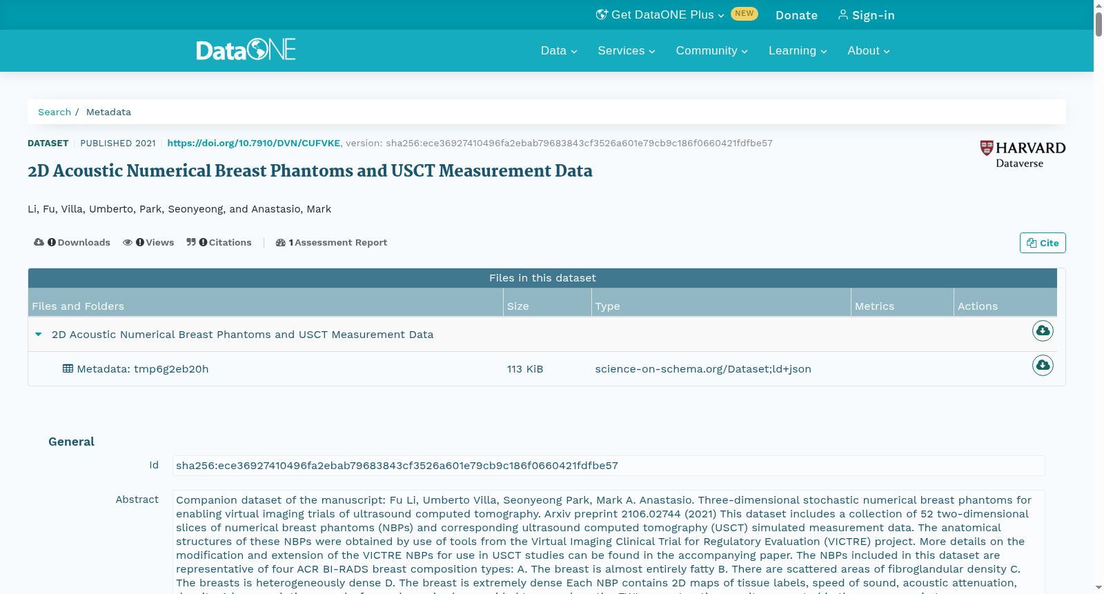
2D Acoustic Numerical Breast Phantoms and USCT Measurement Data
收藏DataONE2021-06-11 更新2024-06-08 收录
下载链接:
https://search.dataone.org/view/sha256:ece36927410496fa2ebab79683843cf3526a601e79cb9c186f0660421fdfbe57
下载链接
链接失效反馈官方服务:
资源简介:
Companion dataset of the manuscript: Fu Li, Umberto Villa, Seonyeong Park, Mark A. Anastasio. Three-dimensional stochastic numerical breast phantoms for enabling virtual imaging trials of ultrasound computed tomography. Arxiv preprint 2106.02744 (2021) This dataset includes a collection of 52 two-dimensional slices of numerical breast phantoms (NBPs) and corresponding ultrasound computed tomography (USCT) simulated measurement data. The anatomical structures of these NBPs were obtained by use of tools from the Virtual Imaging Clinical Trial for Regulatory Evaluation (VICTRE) project. More details on the modification and extension of the VICTRE NBPs for use in USCT studies can be found in the accompanying paper. The NBPs included in this dataset are representative of four ACR BI-RADS breast composition types: A. The breast is almost entirely fatty B. There are scattered areas of fibroglandular density C. The breasts is heterogeneously dense D. The breast is extremely dense Each NBP contains 2D maps of tissue labels, speed of sound, acoustic attenuation, density. A low-resolution speed-of-sound map is also provided to reproduce the FWI reconstruction results presented in the accompanying paper. Corresponding USCT measurement data were simulated by modeling 2D wave propagation in lossy heterogeneous media using a time explicit pseudospectral wave propagation solver. The dataset consists of three folders: The 2d_slices folder contains the 52 slices extracted from 3D NBPs. The measurements folder contains simulated measurement data corresponding to each slice. The imaging_system folder contains information about the 2D imaging system (excitation source, transducer coordinates) In addition, the following helper Matlab scripts are included: read_data.m: Helper function to load and visualize the excitation source and transducer locations. read_waveform_data.m: Helper function to read the .h5 files containing the measurement data. Each slice is saved as a binary MATLAB file (.mat) and contains the following variables label: tissue label map with [2560,2560] pixels and 0.1mm pixel size. Tissue types are denoted using the following labels: water (0), fat (1), skin (2), glandular (29), ligament (88), lesion (200). sos: speed of sound map (mm/μs) with [2560,2560] pixels and 0.1 mm pixel size. Data is stored as data type float32. aa: acoustic attenuation map (Np/m/MHzy) with [2560,2560] pixels and 0.1mm pixel size. Data is stored as data type float32. density: density map (kg/mm3) with [2560,2560] pixels and 0.1 mm pixel size. Data is stored as data type float32. sos_ini: low resolution speed of sound map (mm/μs) with [1280,1280] pixels and 0.2mm pixel size. Data is stored as data type float32. This is the initial guess used in the speed of sound reconstructions in our paper. y: attenuation exponent used for simulation. seed: phantom id type: breast composition type (A-D) The simulated measurement data is saved in hdf5 format. Measurement data corresponding the i-th emitting transducer is stored with hdf5 key equal to the transducer index as a two-dimensional array of size [1024,4250]. Here, the rows represent the receiver index, and the columns the time sample. The sampling frequency is 25MHZ. Because of file size limitations, measurement data for each slice has been divided into 8 chunks, containing data from 128 receivers each. The imaging_system folder contains information regarding the 2D imaging system. source300.mat describes the time profile of the exitation pulse. It consists of 300 time samples at a sampling frequency of 25Mhz. locations1024.mat provide the xy coordinates (mm)of the location of each transducer Data type is float32. Array size is [2x1024]. Warning: This is a very large dataset (~1TB). Please check out our download script written in python.
本数据集为下述论文的配套数据集:Fu Li、Umberto Villa、Seonyeong Park、Mark A. Anastasio,题为《用于超声计算机断层扫描虚拟成像试验的三维随机数值乳房体模》,Arxiv预印本2106.02744(2021)。本数据集包含52组数值乳房体模(Numerical Breast Phantom, NBP)二维切片及其对应的超声计算机断层扫描(Ultrasound Computed Tomography, USCT)模拟测量数据。上述NBP的解剖结构源自监管评估虚拟成像临床试验(Virtual Imaging Clinical Trial for Regulatory Evaluation, VICTRE)项目工具集。针对USCT研究需求修改与扩展VICTRE NBP的更多细节,请参阅配套论文。
本数据集收录的NBP覆盖美国放射学会乳腺影像报告和数据系统(American College of Radiology Breast Imaging Reporting and Data System, ACR BI-RADS)的4种乳腺组织分型:
A. 乳腺几乎完全为脂肪组织
B. 存在散在的纤维腺体密度区域
C. 乳腺为不均匀致密型
D. 乳腺为致密型
每个NBP包含组织标签图、声速(speed of sound, SOS)图、声衰减(acoustic attenuation)图与密度图。此外还提供低分辨率声速图,用于复现配套论文中展示的全波形反演(Full Waveform Inversion, FWI)重建结果。
配套的USCT测量数据通过显式时域伪谱波传播求解器,对有损非均匀介质中的二维波传播进行建模而仿真得到。
本数据集包含三个文件夹:
1. `2d_slices`:包含从3D NBP中提取的52组二维切片
2. `measurements`:包含与每组切片对应的仿真测量数据
3. `imaging_system`:包含二维成像系统的相关信息(激励源、换能器坐标等)
本数据集还附带以下辅助Matlab脚本:
- `read_data.m`:用于加载并可视化激励源与换能器位置的辅助函数
- `read_waveform_data.m`:用于读取存储测量数据的.h5文件的辅助函数
每组切片以二进制MATLAB文件(.mat)格式存储,包含以下变量:
- `label`:组织标签图,分辨率为[2560, 2560]像素,像素尺寸0.1mm。组织类型采用如下标签标注:水(0)、脂肪(1)、皮肤(2)、腺体(29)、韧带(88)、病变(200)
- `sos`:声速图(单位:mm/μs),分辨率为[2560, 2560]像素,像素尺寸0.1mm,数据存储格式为float32
- `aa`:声衰减图(单位:Np/m/MHz),分辨率为[2560, 2560]像素,像素尺寸0.1mm,数据存储格式为float32
- `density`:密度图(单位:kg/mm³),分辨率为[2560, 2560]像素,像素尺寸0.1mm,数据存储格式为float32
- `sos_ini`:低分辨率声速图(单位:mm/μs),分辨率为[1280, 1280]像素,像素尺寸0.2mm,数据存储格式为float32,为本论文中声速重建所用的初始猜测值
- `y`:仿真所用的衰减指数
- `seed`:体模ID
- `type`:乳腺组织分型(A-D)
仿真测量数据以HDF5格式存储。与第i个发射换能器对应的测量数据以换能器索引作为HDF5键值存储,为尺寸[1024, 4250]的二维数组。其中行代表接收换能器索引,列代表时间采样点。采样频率为25MHz。受限于文件大小,每组切片的测量数据被拆分为8个数据块,每个数据块包含128个接收换能器的数据。
`imaging_system`文件夹包含二维成像系统的相关信息:
- `source300.mat`:描述激励脉冲的时域波形,包含300个时间采样点,采样频率为25MHz
- `locations1024.mat`:提供每个换能器的xy坐标(单位:mm),数据存储格式为float32,数组尺寸为[2×1024]
注意:本数据集体量极大(约1TB),请参考我们提供的Python下载脚本进行下载。
创建时间:
2023-11-19
搜集汇总
数据集介绍
以上内容由遇见数据集搜集并总结生成


