<b>Supplemental Material</b>:<b>Supplemental Figures</b> <b>S1-S5, </b><b>Supplemental Videos S1-S5</b>
收藏资源简介:
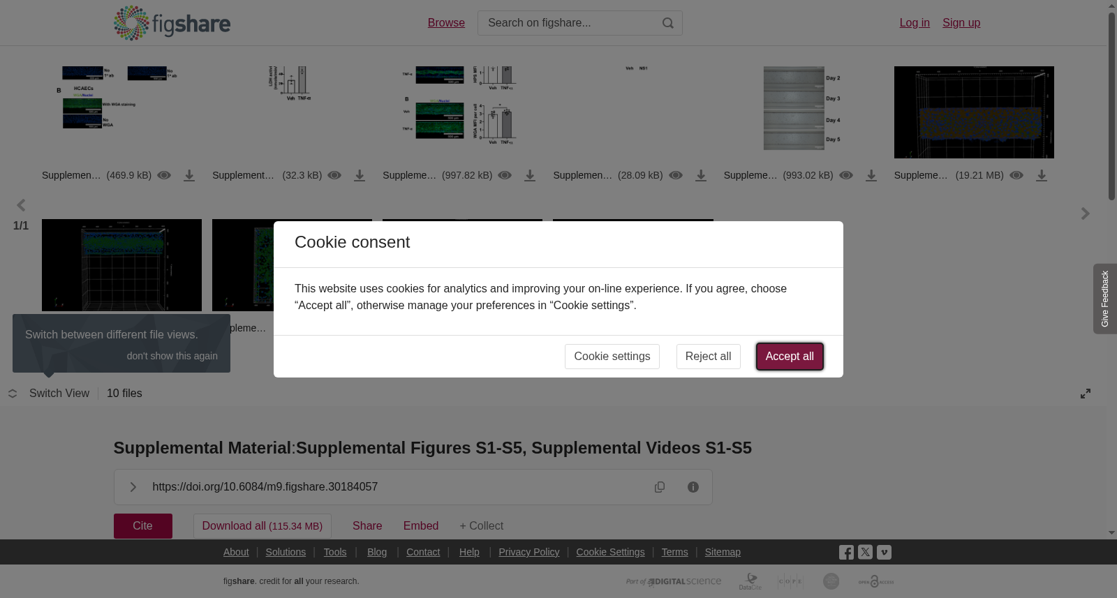
<b>Supplemental Figure S1. </b>Representative images for (A) primary antibody (1o ab) validation of heparan sulfate (HS) and (B) for wheat germ agglutinin (WGA) staining in 3D endothelium. Human umbilical vein endothelial cells (HUVECs) and human coronary artery endothelial cells (HCAECs). Magnification: ×10; scale bar: 500 μm, (n=2-3).<b>Supplemental Figure S2.</b> Cytotoxicity effect of TNF-α on 3D endothelium. Lactate dehydrogenase (LDH) activity in 3D HUVECs following treatment of either vehicle (Veh) or 10 ng/ml TNF-α for 24 h. Statistical analysis was determined by Student’s t-test (*p<0.05 vs. Veh). Data are expressed as mean ± SD (n=3-5).<b>Supplemental Figure S3. </b>Representative fluorescence images and quantification of mean fluorescence intensity (MFI) per cell of (A) heparan sulfate (HS) and (B) EGX components stained with wheat germ agglutinin (WGA) after 3D HUVECs were stimulated with Veh or 1 ng/mL TNF-α for 5 h. Magnification: ×10; scale bar: 500 μm. Student’s t-test was used to compare Veh and TNF-α (*p < 0.05 vs Veh; ns = not significant). Data are presented as means ± SDs (n = 8).<br><b>Supplemental Figure S4.</b> Cytotoxicity effects of NS1 on 3D endothelium. Lactate dehydrogenase (LDH) activity in 3D HUVECs treated with vehicle (Veh) or NS1 for 12 h. Statistical analysis was determined via Student’s<i> t</i>-test (ns = not significant). Data are presented as means ± SDs (n = 7).<b>Supplemental Figure S5</b><b>.</b> Representative bright-field images of 3D HCAECs from Day 0 to Day 5. HCAECs were cultured in two-lane Organoplates (Mimetas), forming 3D tubular structures. Magnification: ×5; scale bar: 200 μm.<br><b>Supplemental Video S1.</b> Characterization of 3D endothelium. Human umbilical vein endothelial cells (HUVECs) were cultured in two-lane Organoplates (Mimetas), forming 3D tubular structures within 4–5 days. The 3D HUVECs were stained for CD31 (green), F-actin (red), and nuclei (blue). The 3D architecture was visualized using Z-plane images captured at 8-μM intervals in confocal mode with the Opera Phenix High-Content Screening System.<br><b>Supplemental Video S2 and S3. </b>Comparisons of heparan sulfate (HS) expression between 2D endothelial cell cultures and 3D endothelium. Human umbilical vein endothelial cells (HUVECs) were cultured in <b>(</b><b>S2</b><b>) </b>24-well plates until 95% confluency or <b>(</b><b>S3</b><b>)</b> two-lane Organoplates to generate 3D endothelium. They were stained for HS (green) and nuclei (blue). Images were captured using Z-plane images at 8-μM intervals in confocal mode via the Opera Phenix High-Content Screening System.<br><b>Supplemental Video S4 and S5. </b>Comparisons of wheat germ agglutinin (WGA) staining between 2D endothelial cell cultures and 3D endothelium. Human umbilical vein endothelial cells (HUVECs) were cultured in <b>(</b><b>S4</b><b>)</b> 24-well plates until 95% confluency or <b>(</b><b>S5</b><b>)</b> two-lane Organoplates to generate 3D endothelium. They were stained with WGA (green) and nuclei (blue). Images were captured using Z-plane images at 8-μM intervals in confocal mode via the Opera Phenix High-Content Screening System.
**补充图S1。** 用于(A)硫酸乙酰肝素(heparan sulfate, HS)一抗(primary antibody, 1o ab)验证,以及(B)3D内皮模型中小麦凝集素(wheat germ agglutinin, WGA)染色的代表性图像。实验采用人脐静脉内皮细胞(human umbilical vein endothelial cells, HUVECs)与人冠状动脉内皮细胞(human coronary artery endothelial cells, HCAECs)。放大倍数:×10;标尺:500 μm,样本量n=2-3。 **补充图S2。** 肿瘤坏死因子α(tumor necrosis factor-α, TNF-α)对3D内皮模型的细胞毒性作用。分别以溶剂对照(vehicle, Veh)或10 ng/ml TNF-α处理3D人脐静脉内皮细胞24小时后,检测其乳酸脱氢酶(lactate dehydrogenase, LDH)活性。统计学分析采用学生t检验(Student’s t-test)(*p<0.05 与Veh组相比)。数据以平均值±标准差(mean ± SD)表示,样本量n=3-5。 **补充图S3。** 3D人脐静脉内皮细胞经溶剂对照或1 ng/mL TNF-α刺激5小时后,(A)硫酸乙酰肝素(HS)及(B)EGX组分经小麦凝集素(WGA)染色的代表性荧光图像及每细胞平均荧光强度(mean fluorescence intensity, MFI)定量结果。放大倍数:×10;标尺:500 μm。采用学生t检验(Student’s t-test)比较溶剂对照组与TNF-α处理组(*p < 0.05 与Veh组相比;ns = 无显著性差异)。数据以平均值±标准差(means ± SDs)表示,样本量n=8。 **补充图S4。** NS1对3D内皮模型的细胞毒性作用。以溶剂对照(Veh)或NS1处理3D人脐静脉内皮细胞12小时后,检测其乳酸脱氢酶(LDH)活性。统计学分析采用学生t检验(Student’s t-test)(ns = 无显著性差异)。数据以平均值±标准差(means ± SDs)表示,样本量n=7。 **补充图S5。** 第0天至第5天的3D人冠状动脉内皮细胞明场代表性图像。人冠状动脉内皮细胞于双通道Organoplates(Mimetas)中培养,形成3D管状结构。放大倍数:×5;标尺:200 μm。 **补充视频S1。** 3D内皮模型的表征。将人脐静脉内皮细胞(HUVECs)接种于双通道Organoplates(Mimetas)中,于4~5天内形成3D管状结构。对3D人脐静脉内皮细胞进行CD31(绿色)、F-肌动蛋白(F-actin)及细胞核(蓝色)染色。采用Opera Phenix高内涵筛选系统(Opera Phenix High-Content Screening System)以共聚焦模式采集间隔8 μm的Z层图像,以可视化3D结构。 **补充视频S2与S3。** 二维(2D)内皮细胞培养与三维(3D)内皮模型中硫酸乙酰肝素(HS)表达的比较。将人脐静脉内皮细胞(HUVECs)分别于(S2)24孔板中培养至汇合度95%,或(S3)双通道Organoplates中培养以构建3D内皮模型。随后对其进行HS(绿色)及细胞核(蓝色)染色。采用Opera Phenix高内涵筛选系统以共聚焦模式采集间隔8 μm的Z层图像。 **补充视频S4与S5。** 二维(2D)内皮细胞培养与三维(3D)内皮模型中小麦凝集素(WGA)染色的比较。将人脐静脉内皮细胞(HUVECs)分别于(S4)24孔板中培养至汇合度95%,或(S5)双通道Organoplates中培养以构建3D内皮模型。随后用WGA(绿色)及细胞核(蓝色)进行染色。采用Opera Phenix高内涵筛选系统以共聚焦模式采集间隔8 μm的Z层图像。