
Clinical and Histopathological Evaluation of a Rabbit Model for <i>Pythium insidiosum</i> Keratitis
收藏DataCite Commons2020-08-25 更新2024-07-28 收录
下载链接:
https://tandf.figshare.com/articles/Clinical_and_Histopathological_Evaluation_of_a_Rabbit_Model_for_i_Pythium_insidiosum_i_Keratitis/11919927
下载链接
链接失效反馈官方服务:
资源简介:
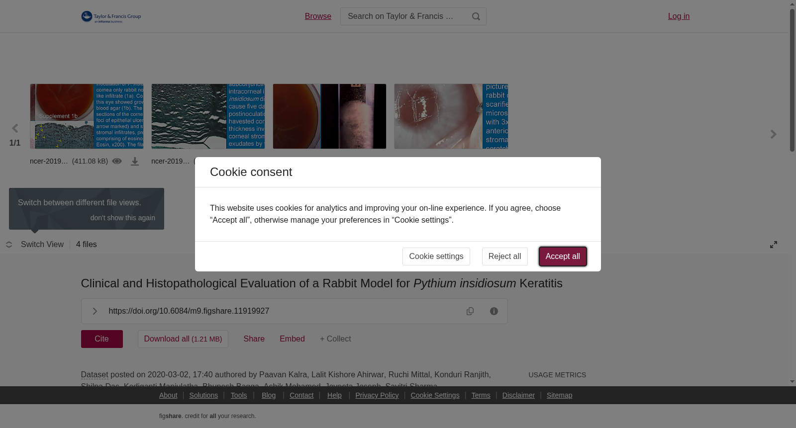
<b>Purpose</b>: To describe the clinical characteristics and histopathological features in a rabbit model of <i>Pythium insidiosum</i> keratitis. <b>Methods</b>: Zoospores of <i>P. insidiosum</i> isolated from a patient with microbial keratitis were used for inoculation of the right eye of 48 <i>New Zealand White</i> rabbits in either low (LD) or high dose (HD). Apart from variable dosage the rabbits were grouped (6 rabbits per group) based on route of inoculation (topical on abraded cornea or intracorneal) and immunosuppression (subconjunctival steroid or no steroid). Left eye received phosphate buffered saline via route similar to the right eye. Daily clinical examination of the eye was done, the corneas were harvested on day 3, 7 and 9 and part of the cornea was preserved in 10% neutral buffered formalin for histopathological examination. <b>Results</b>: Left eye of all rabbits were clinically normal. Eyes with intracorneal injection of zoospores developed infection irrespective of dose of inoculation and administration of steroids. One of the consistent early signs of infection was ring like infiltrate in the peripheral cornea. On day 2, rabbits receiving HD developed significantly greater inflammation compared to LD [median clinical score in HD- 11 (IQR = 10–12), versus 9 (IQR = 8–9) in LD (<i>p</i> = 0.004)]. The density of inflammation showed temporal correlation (increase with time) when the inoculum was low. Of the rabbits that received topical inoculation one rabbit cornea showed mild infiltrate in steroid group while no eye was infected in the group without steroid. Sparsely septate to aseptate branching filaments were noted in the stroma of all infected corneas. <b>Conclusions</b>: We describe the first animal model of <i>Pythium</i> keratitis that holds promise for future studies. While topical inoculation of zoospores was unsuccessful in causing infection intracorneal inoculation without immunosuppression was sufficient to develop clinically severe keratitis in rabbits.
【研究目的】本研究旨在描述隐秘腐霉(Pythium insidiosum)性角膜炎兔模型的临床特征与组织病理学特征。【研究方法】本研究使用从1例微生物性角膜炎患者体内分离得到的隐秘腐霉游动孢子,以低剂量(LD)或高剂量(HD)接种48只新西兰白兔(New Zealand White)的右眼。除接种剂量存在差异外,实验兔按接种途径(角膜擦伤后局部滴注或角膜内注射)及免疫抑制方案(结膜下注射糖皮质激素或不使用糖皮质激素)分为6组,每组6只。家兔左眼以与右眼完全一致的途径接种磷酸盐缓冲盐水作为对照。每日对家兔眼部进行临床检查,分别于接种后第3、7、9天摘取角膜,取部分角膜组织置于10%中性缓冲福尔马林中固定,用于组织病理学检测。【研究结果】所有家兔左眼均未出现临床异常表现。无论接种剂量与糖皮质激素使用情况如何,经角膜内注射游动孢子的眼部均发生感染。感染早期的一致性体征之一为周边角膜出现环状浸润灶。接种后第2天,高剂量组家兔的炎症反应显著高于低剂量组[高剂量组中位临床评分为11(四分位距IQR=10~12),低剂量组为9(IQR=8~9),p=0.004]。当接种剂量较低时,炎症密度随时间推移逐渐升高,呈显著时间相关性。在接受局部接种的家兔中,糖皮质激素处理组仅1只家兔角膜出现轻度浸润,而未使用糖皮质激素的组别未发生眼部感染。所有受感染角膜的基质层中均可见稀疏分隔至无分隔的分支菌丝。【研究结论】本研究首次构建了隐秘腐霉性角膜炎动物模型,可为后续相关研究提供可靠的实验基础。尽管局部接种游动孢子未能成功诱导感染,但不使用免疫抑制剂的角膜内接种即可使家兔出现临床意义上的重度角膜炎。
提供机构:
Taylor & Francis创建时间:
2020-03-02
搜集汇总
数据集介绍
背景与挑战
背景概述
该数据集描述了兔子模型用于Pythium insidiosum角膜炎的临床和组织病理学评估研究。通过接种游动孢子到48只新西兰白兔,分组考察剂量、接种途径和免疫抑制的影响,结果显示角膜内注射能有效引发感染,并首次建立了该疾病的动物模型,为未来研究提供了基础。
以上内容由遇见数据集搜集并总结生成

暂无相关数据集


