Original data-In situ observations of gold deposition in a dense liquid layer at the pyrite-water interface
收藏资源简介:
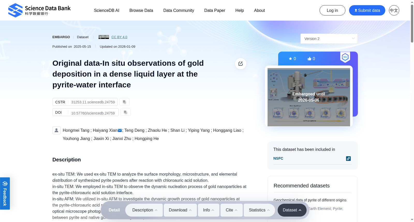
ex-situ TEM: We used ex-situ TEM to analyze the surface morphology, microstructure, and elemental distribution of synthesized pyrite powders after reaction with chloroauric acid solution.in-situ TEM: We employed in-situ TEM to observe the dynamic nucleation process of gold nanoparticles at the pyrite-chloroauric acid solution interface.in-situ AFM: We utilized in-situ AFM to investigate the dynamic growth process of gold nanoparticles at the pyrite-chloroauric acid solution interface.optical microscope photographs: We employed optical microscopy to examine the spatial relationship between pyrite and native gold in Jinshan gold deposit.SEM+XRD: We used XRD and SEM to analyze the crystal structure and micro-morphology of synthesized pyrite powders.XPS: We utilized XPS to detect the chemical environment evolution on pyrite surfaces after in-situ AFM reactions.These data provide critical insights into the in-situ nucleation and growth processes of gold nanoparticles at the pyrite-water interface, significantly advancing our understanding of gold mineralization mechanisms.
离线透射电子显微镜(ex-situ TEM):本研究采用离线透射电子显微镜(ex-situ TEM),对与氯金酸溶液反应后的合成黄铁矿粉末的表面形貌、微观结构及元素分布开展分析。 原位透射电子显微镜(in-situ TEM):本研究借助原位透射电子显微镜(in-situ TEM),实时观测黄铁矿-氯金酸溶液界面处金纳米颗粒的动态成核过程。 原位原子力显微镜(in-situ AFM):通过原位原子力显微镜(in-situ AFM),探究黄铁矿-氯金酸溶液界面处金纳米颗粒的动态生长过程。 光学显微镜成像:采用光学显微技术,考察金山金矿床中黄铁矿与自然金的空间分布关系。 扫描电子显微镜(SEM)与X射线衍射(XRD)联用:结合X射线衍射(XRD)与扫描电子显微镜(SEM),分析合成黄铁矿粉末的晶体结构与微观形貌。 X射线光电子能谱(XPS):利用X射线光电子能谱(XPS),检测经原位原子力显微镜反应后黄铁矿表面的化学环境演化。 本系列实验数据为阐明黄铁矿-水界面处金纳米颗粒的原位成核与生长过程提供了关键依据,显著深化了对金成矿机制的认知。