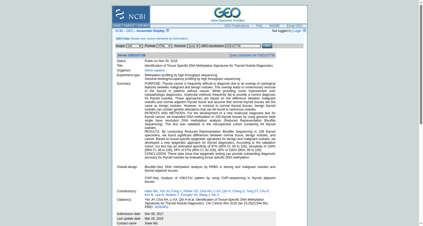
Identification of Tissue Specific DNA Methylation Signatures for Thyroid Nodule Diagnostics
收藏资源简介:
PURPOSE: Thyroid cancer is frequently difficult to diagnose due to an overlap of cytological features between malignant and benign nodules. This overlap leads to unnecessary removal of the thyroid in patients without cancer. While providing some improvement over cytopathologic diagnostics, molecular methods frequently fail to provide a correct diagnosis for thyroid nodules. These approaches are based on the difference between malignant nodules and normal adjacent thyroid tissue and assume that normal thyroid tissues are the same as benign nodules. However, in contrast to normal thyroid tissues, benign thyroid nodules can contain genetic alterations that can be found in cancerous nodules. PATIENTS AND METHODS: For the development of a new molecular diagnostic test for thyroid cancer, we evaluated DNA methylation in 109 thyroid tissues by using genome wide single base resolution DNA methylation analysis (Reduced Representation Bisulfite Sequencing). The test was validated in the retrospective cohort containing 64 thyroid nodules. RESULTS: By conducting Reduced Representation Bisulfite Sequencing in 109 thyroid specimens, we found significant differences between normal tissue, benign nodules, and cancer. Based on tissue-specific epigenetic signatures for benign and malignant nodules, we developed a new epigenetic approach for thyroid diagnostics. According to the validation cohort, our test has an estimated specificity of 97% (95% CI, 80 to 100), sensitivity of 100% (95% CI, 86 to 100), PPV of 97% (95% CI, 82-100), NPV of 100% (95%, 85 to 100). CONCLUSION: These data show that epigenetic testing can provide outstanding diagnostic accuracy for thyroid nodules by evaluating tissue specific DNA methylation. Bisulfite-Seq: DNA methylation analysis by RRBS in bening and malignant nodules and thyroid adjacent tissues ChIP-Seq: Analysis of H3K27Ac pattern by using ChIP-sequencing in thyroid adjacent tissues
研究背景:甲状腺癌的良恶性结节细胞学特征存在重叠,常导致诊断困难,进而对未患癌症的患者实施不必要的甲状腺切除术。现有分子诊断方法虽较细胞病理学诊断有所进步,但往往仍无法对甲状腺结节作出准确诊断——这类方法基于恶性结节与邻近正常甲状腺组织的差异,且默认正常甲状腺组织与良性结节完全一致。然而,与正常甲状腺组织不同的是,良性甲状腺结节可携带癌结节中存在的遗传改变。 研究对象与方法:为开发新型甲状腺癌分子诊断检测方法,我们采用全基因组单碱基分辨率DNA甲基化分析技术——简化代表性亚硫酸氢盐测序(Reduced Representation Bisulfite Sequencing, RRBS),对109份甲状腺组织的DNA甲基化水平进行了检测。本检测在包含64枚甲状腺结节的回顾性队列中完成验证。 研究结果:通过对109份甲状腺标本开展简化代表性亚硫酸氢盐测序,我们发现正常组织、良性结节与癌组织之间存在显著差异。基于良恶性结节的组织特异性表观遗传特征,我们开发了一种新型甲状腺诊断表观遗传学检测方法。经验证队列分析,本检测的特异性约为97%(95%置信区间:80~100),灵敏度为100%(95%置信区间:86~100),阳性预测值(Positive Predictive Value, PPV)为97%(95%置信区间:82~100),阴性预测值(Negative Predictive Value, NPV)为100%(95%置信区间:85~100)。 研究结论:本研究数据表明,通过评估组织特异性DNA甲基化水平,表观遗传学检测可为甲状腺结节诊断提供极高的诊断准确性。 附加测序分析说明: 1. 亚硫酸氢盐测序(Bisulfite-Seq):针对良性、恶性结节及甲状腺邻近组织,采用RRBS开展DNA甲基化分析; 2. 染色质免疫共沉淀测序(ChIP-Seq):在甲状腺邻近组织中采用ChIP测序分析H3K27Ac修饰模式。